Get Care
Hydatid Cyst
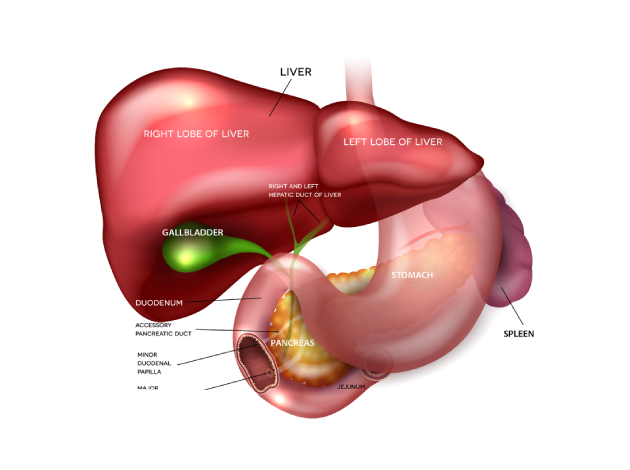
What is a Hydatid Cyst?
A hydatid cyst is a rare, parasitic infection caused by the tapeworm Echinococcus. It primarily affects the liver, but can also involve other organs, including the lungs, brain, and bones. The cysts form as the larval stage of the tapeworm, which grows and produces fluid-filled sacs. These sacs can grow larger over time, leading to potential complications depending on the location and size of the cysts.
Humans can become infected with Echinococcus by ingesting the tapeworm eggs, often through contact with infected animals such as dogs, sheep, or cattle, or through consumption of contaminated food or water. Although the infection is more common in regions where livestock farming is prevalent, it can occur anywhere in the world.
What Causes Hydatid Cyst?
Hydatid cysts are caused by infection with the Echinococcus tapeworm, leading to a condition called hydatid disease or cystic echinococcosis. The parasite primarily affects animals but can accidentally infect humans.
Lifecycle and Transmission
The Echinococcus parasite has two hosts:
Definitive Hosts
Carnivores like dogs, wolves, and foxes carry the adult tapeworms in their intestines. They pass eggs in their feces, contaminating soil, water, and plants.Intermediate Hosts
Herbivores such as sheep, goats, and cattle ingest these eggs while grazing. The eggs hatch into larvae, which travel through the bloodstream and form hydatid cysts in organs—mainly the liver and lungs.
The cycle continues when a carnivore eats infected organs of an intermediate host.
How Humans Get Infected (Accidental Hosts)
Humans may become accidental hosts by:
Ingesting eggs through contaminated food, water, or soil.
Contact with infected dogs or wild animals (especially feces or fur).
Poor hygiene after handling animals or their environment.
Once ingested, the eggs hatch in the intestine, and larvae spread to the liver, lungs, or other organs, where they form slow-growing fluid-filled cysts. These cysts can cause pressure symptoms, allergic reactions, or complications if ruptured.
What are the Symptoms of Hydatid Cyst?
The symptoms of a hydatid cyst depend on the size and location of the cyst. Some people may remain asymptomatic for years, while others may experience symptoms when the cyst grows large enough to cause pressure on surrounding tissues or organs.
Common symptoms include:
- Abdominal Pain: If the cyst is located in the liver or spleen, it can cause dull, aching pain in the upper right abdomen.
- Jaundice: A hydatid cyst in the liver can block bile ducts, leading to jaundice (yellowing of the skin and eyes).
- Fever: The infection may cause low-grade fever, particularly if the cyst becomes infected or ruptures.
- Nausea and Vomiting: These symptoms may occur if the cyst presses on the stomach or intestines.
- Chest Pain or Difficulty Breathing: If the cyst is located in the lungs, it may cause chest discomfort, shortness of breath, or a persistent cough.
- Coughing up Blood: A ruptured cyst in the lungs can lead to coughing up blood or a blood-tinged sputum.
- Fatigue: Chronic infection or complications from the cyst may lead to fatigue and general weakness.
- Anaphylactic Shock: In rare cases, if a cyst ruptures, it can cause an allergic reaction, leading to severe symptoms such as difficulty breathing, a drop in blood pressure, and swelling.
How is Hydatid Cyst Diagnosed?
Diagnosis of a hydatid cyst involves a combination of medical history, physical examination, and various diagnostic tests. If a hydatid cyst is suspected, the following tests may be used:
- Imaging Tests:
- Ultrasound: This is the most common test to detect hydatid cysts. It can reveal the characteristic fluid-filled cysts in the liver, lungs, or other organs.
- CT Scan (Computed Tomography): A CT scan provides detailed cross-sectional images of the organs and can help determine the size, location, and number of cysts. It is particularly useful for detecting cysts in the lungs or brain.
- MRI (Magnetic Resonance Imaging): MRI is used for more detailed imaging of the brain and spinal cord if the cysts are suspected to be in these areas.
- Blood Tests:
- Serologic Tests: Blood tests can detect antibodies against Echinococcus. These tests are useful for diagnosing hydatid disease, especially in cases where cysts are located in organs that are hard to image, such as the brain.
- Eosinophil Count: A high eosinophil count in the blood may indicate a parasitic infection like hydatid cysts.
- Biopsy: In rare cases, a biopsy may be performed to obtain a sample of the cyst tissue for laboratory analysis. This is usually done when non-invasive tests are inconclusive or when the cyst needs to be examined further for potential complications.
What are the Treatment Options for Hydatid Cyst?
The treatment of hydatid cysts depends on the size, location, and symptoms of the cyst, as well as the overall health of the patient. Common treatment approaches include:
- Surgery:
Surgical removal of the cyst is the most common treatment, particularly for large or symptomatic cysts. The surgery involves carefully removing the cyst from the affected organ, often with a surrounding layer of healthy tissue to prevent leakage of cyst fluid, which can cause anaphylaxis or spread the infection. In some cases, the cyst may be drained before removal. For cysts in the liver, a partial hepatectomy (removal of part of the liver) may be necessary. - Antiparasitic Medications:
In some cases, especially for smaller or asymptomatic cysts, antiparasitic medications such as albendazole or mebendazole may be prescribed to help kill the tapeworm larvae inside the cysts. These medications are often used in conjunction with surgery to prevent recurrence of the infection. - Percutaneous Aspiration (Needle Drainage):
In certain cases, a needle may be inserted into the cyst to aspirate (drain) the fluid. This can help reduce the size of the cyst or alleviate symptoms. However, this method carries the risk of causing the cyst to rupture or spread the infection, so it is used with caution. - Laparoscopic Surgery:
In some cases, laparoscopic (minimally invasive) surgery can be used to remove the cyst. This approach involves smaller incisions and results in less recovery time and fewer complications compared to traditional open surgery. - Post-Surgical Care:
After surgery, patients will need regular follow-up visits to monitor for any signs of recurrence. Antiparasitic medications may continue for several weeks to ensure that any remaining larvae are eradicated.
What are the Complications of Hydatid Cyst?
If left untreated, hydatid cysts can lead to several serious complications:
- Cyst Rupture: The cyst may rupture, releasing cyst fluid and larvae into the body, which can cause a severe allergic reaction (anaphylaxis), infection, or the spread of the parasite to other organs.
- Infection: If the cyst becomes infected, it can lead to abscess formation, sepsis, and other life-threatening complications.
- Organ Dysfunction: Large cysts can damage the organ in which they are located, such as the liver or lungs, leading to organ failure or impairment of function.
- Recurrence: If the cyst is not completely removed or treated, there is a risk of the infection returning, potentially causing further complications.
Why Choose Us?
Contact Us